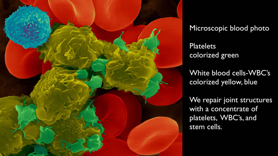
In this newsletter, let’s look at the PRP Stem Cell Joint Repair process through a microscope.
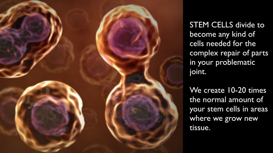
We concentrate platelets, white blood cells, and stem cells from your blood.
text added on this website
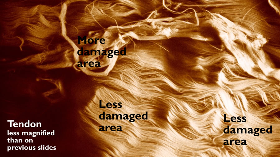
One common target is tendon.
text added on this website
The strands in the above picture are fibers. Each fiber is composed of many smaller threads called fibrils.
text added on this website
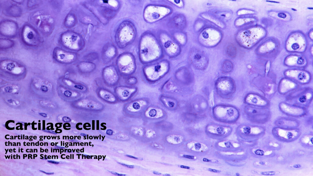
Yet another target of our injections is cartilage.
text added on this website
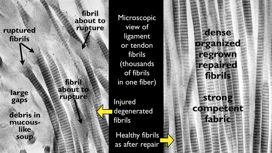
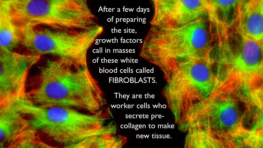
Once the platelet and stem cell concentrate touches tendon, ligament, or cartilage, a complex and glorious repair process begins. One step in that process is that certain white blood cells called fibroblasts develop in the injected areas of wear and tear. They secrete a precursor to collagen that combines with Vitamin C in your body to make new repair collagen.
Dept. of Cell & Developmental Biology, University of North Carolina-Charlotte
text added on this website
I’ll share a tune called A Closer View, by Gary Peacock and Ralph Towner:
After all the above zooming in, I’m going to zoom out. When I was younger, my father said I had my head in the clouds. He was right, as evidenced by my fascination with photographing clouds and the sky. I share some examples below.
Enjoy having your head in the clouds.
Dr. Jonas Skardis